
A mixed chest-imaging result
Li Shuying · 74 years · Inner Mongolia
Source image 01Open full resolution ↗
Read the English image text
MYYL
Lung imaging follow-up
PATIENT FOLLOW-UP RECORDS
Li Shuying · Female · 74 · Inner Mongolia
English translation · Original medical images retained
BASELINE · Symptoms and CT report
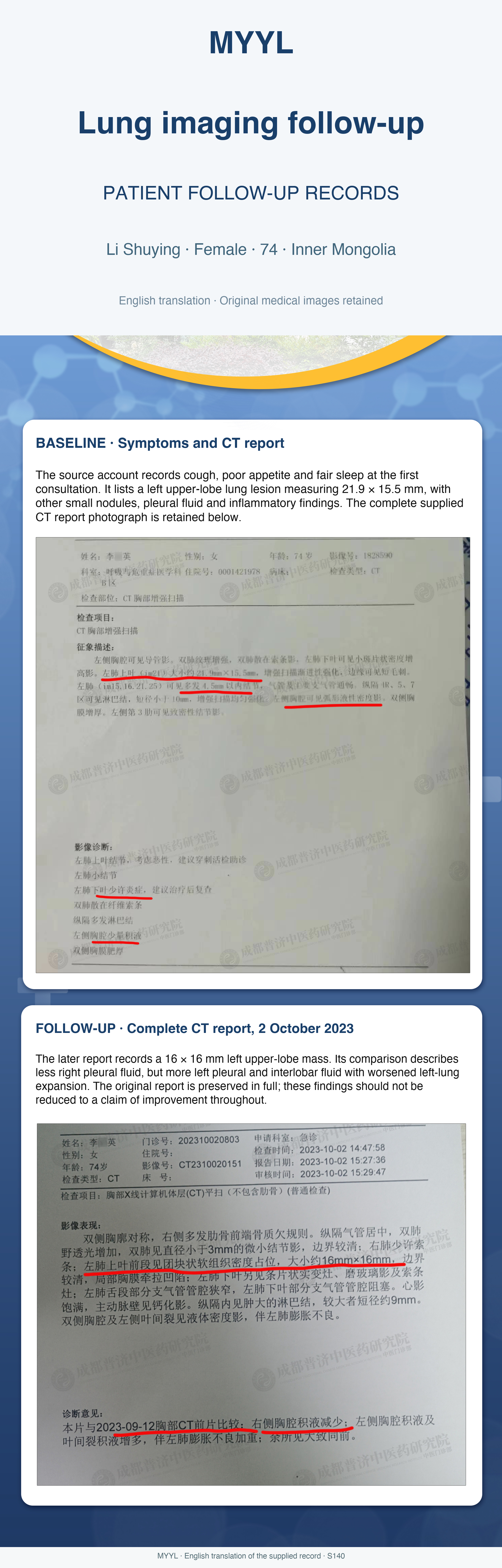
The source account records cough, poor appetite and fair sleep at the first consultation. It lists a left upper-lobe lung lesion measuring 21.9 × 15.5 mm, with other small nodules, pleural fluid and inflammatory findings. The complete supplied CT report photograph is retained below.
FOLLOW-UP · Complete CT report, 2 October 2023
The later report records a 16 × 16 mm left upper-lobe mass. Its comparison describes less right pleural fluid, but more left pleural and interlobar fluid with worsened left-lung expansion. The original report is preserved in full; these findings should not be reduced to a claim of improvement throughout.
English report translation
MYYL | S140 | English translation of visible report text
Both complete supplied CT report photographs are preserved unchanged in the accompanying image. These are written reports; the source does not contain CT slice images.
FIRST REPORT — CONTRAST-ENHANCED CHEST CT
Patient: Female, 74 years. Examination date is not visible in the supplied photograph.
Findings: A catheter is visible in the left pleural cavity. Lung markings are increased. Scattered fibrous bands are seen in both lungs, with a small patchy increased-density area in the left lower lobe. A left upper-lobe lesion measures approximately 21.9 × 15.5 mm, with progressive enhancement and short marginal spicules. Several small left-lung nodules measure up to 4.5 mm. The trachea and bronchial lumens are patent. Mediastinal lymph nodes are visible with short axes below 10 mm and homogeneous enhancement. A small amount of fluid is present in the left pleural cavity. Both pleura are thickened. A dense nodular focus is visible in the left third rib.
Impression: Left upper-lobe nodule, considered suspicious for malignancy; biopsy is suggested to assist diagnosis. Small left-lung nodules. A small inflammatory focus in the left lower lobe; follow-up after treatment is suggested. Scattered fibrous bands in both lungs. Multiple mediastinal lymph nodes. Small left pleural effusion. Bilateral pleural thickening.
SECOND REPORT — NON-CONTRAST CHEST CT
Examination: 2 October 2023, 14:47:58. Report: 2 October 2023, 15:27:36.
Findings: The chest is symmetrical. The anterior ends of several right ribs show some bony irregularity. The mediastinum and trachea are central. Lung lucency is increased. Both lungs show tiny nodules below 3 mm, with relatively clear boundaries. A few linear bands are present in the right lung. A soft-tissue mass in the anterior segment of the left upper lobe measures approximately 16 × 16 mm, with a relatively clear boundary and local pleural retraction. Additional areas of consolidation, ground-glass opacity and linear bands are present in the left lower lobe. Some bronchi in the lingula are narrowed and some in the left lower lobe are obstructed. The cardiac silhouette is full; aortic-wall calcification is present. Mediastinal lymph nodes are enlarged, the larger approximately 9 mm in short axis. Fluid-density areas are seen in both pleural cavities and the left interlobar fissure, with incomplete expansion of the left lung.
Comparison with the chest CT of 12 September 2023: Right pleural fluid has decreased. Left pleural and interlobar fluid have increased, with worsening incomplete left-lung expansion. Other findings are broadly unchanged.
Source note: The date of the first photographed report is not visible, so it cannot automatically be identified as the 12 September comparison study. The later report describes mixed changes.
More lung cancer records
 1 source image
Lung cancer
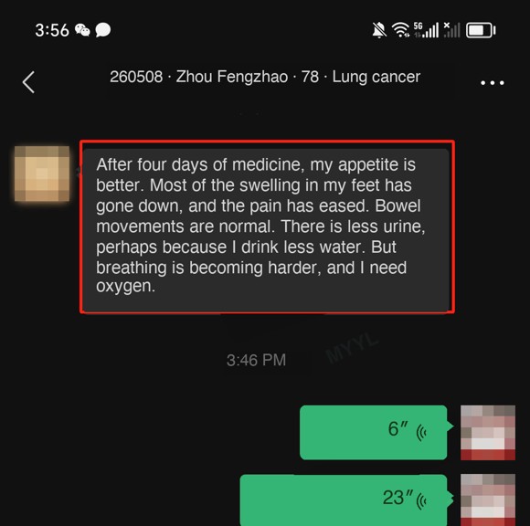
Appetite improved; breathing became harder
After four days of medication, the correspondent reported a better appetite, less swelling in the feet and some pain relief.
 2 source images
Lung cancer
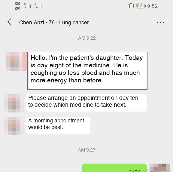
Two updates on coughing up blood
On day eight of medication, Chen's daughter said coughing up blood had become less frequent and his energy had improved.
 1 source image
Lung cancer
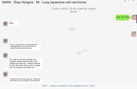
Less arm pain, with stomach discomfort continuing
On day six of medication, Zhao said abdominal bloating had eased but her stomach still felt uncomfortable.